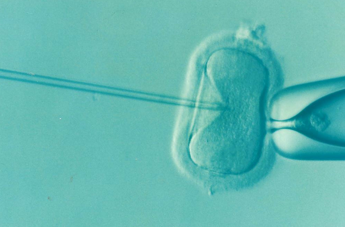
The process of in vitro fertilization (IVF) is often unsuccessful due to chromosomal changes that occur in an embryo fertilized in a test tube. So far, it was not known whether these changes also transfer to the baby, but an article published in the Nature Medicine journal shows that these genetically mutated cell lines are not inherited to the child.
Compared to the mother’s body, the egg fertilized in a test tube has rather different conditions for growing. “As these conditions are not consistent with those the embryo in a woman’s organism has, this creates a situation where chromosomal changes occur in the embryo quite often, referred as embryo mosaicism” said Dr Andres Salumets, a professor of reproductive medicine at the University of Tartu and Head of the Competence Centre on Health Technologies.
After fertilization, the fertilized oocyte or a zygote starts to rapidly develop, which generates many genetic abnormalities in some or all embryonal cells. These errors are caused by a process named chromosomal instability.
When the IVF embryo is transferred into the mother’s body, it is often mosaic, i.e., simultaneously containing chromosomally normal as well as abnormal cells. Prior studies have shown that similar mosaicism may also occur in naturally conceived children, but very likely the mosaicism of IVF embryos is more common. This has given rise to hot discussions: could it pose a genetic risk to the child to be born?
“Even when using very sensitive methods, we could not see any cell lines with chromosomal aberrations in IVF children. Thus, our study shows that in vitro fertilization does not pose any health risks to the children to be born,” explained professor Salumets.
In addition to the knowledge of geneticists, this kind of an interdisciplinary study required a cooperation with infertility clinicians, clinical embryologists and bioinformaticians. The current study became possible via an advanced computational method, called haplarithmisis. This enabled to dissect the cellular composition of placenta and estimate maternal and foetal compartments of the placenta, allowing identification of mosaic genetic imbalances in unprecedented detail in both the child’s own genome and the placental tissue.
Scientists looked at live-born children. “The implications of chromosomal instability in difficult pregnancies are still to be studied, including preeclampsia and small-for-gestational age children that are caused by placenta malfunction,” said Dr Salumets.
The study published in the Nature Medicine journal was the result of a four-year cooperation by Estonian, Belgian, Dutch and Finnish scientists.
The translation of this article from Estonian Public Broadcasting science news portal Novaator was funded by the European Regional Development Fund through Estonian Research Council.
Photo by: Pixabay.com